
Radiation has been an effective tool for treating cancer for more than 100 years. More than 60 percent of patients diagnosed with cancer will receive radiation therapy as part of their treatment.
Radiation therapy works by damaging the DNA of cells and destroys their ability to reproduce. Both normal and cancer cells can be affected by radiation, but cancer cells have generally impaired ability to repair this damage, leading to cell death.
Therapeutic radiation serves two major functions.To cure cancer: Destroy tumors that have not spread. Kill residual microscopic disease left after surgery or chemotherapy.To reduce or palliate symptoms: Shrink tumors affecting quality of life, e.g., a lung tumor causing shortness of breath. Alleviate pain or neurologic symptoms by reducing the size of a tumor.
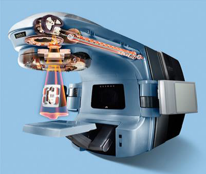 Fig 1:
Most external beam radiation treatments use photons
The delivery of external beam radiation treatment is painless and usually scheduled five days a week for several weeks.
Clinical or Radiation Oncologist: The doctor who prescribes and oversees the radiation therapy treatments.Medical Physicist: Ensures that treatment plans are properly tailored for each patient, and is responsible for the calibration and accuracy of treatment equipment. Dosimetrist: Works with the oncologist and medical physicist to calculate the proper dose of radiation given to the tumor.Radiation Therapist: Administers the daily radiation under the doctors prescription and supervision. Oncology Nurse: Interacts with the patient and family at the time of consultation, throughout the treatment process and during follow-up care.
 Fig 2:
DNA Double Helix
Referral, Consultation, Simulation, Treatment Planning, Quality Assurance.
Patient is set up in treatment position on a dedicated CT scanner. Immobilization devices used to create to assure patient comfort and daily reproducibility.Reference marks or tattoos may be placed on patient.CT simulation images are often fused with PET or MRI scans for treatment planning.
Physician outlines/contour the target and organs at risk. Sophisticated software are used to carefully derive an appropriate treatment plan. Computerized algorithms enable the treatment plan to spare as much healthy tissue as possible. Medical physicist checks the chart and dose calculations. Oncologist reviews and approves final plan.
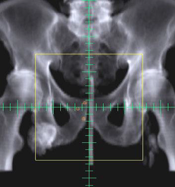 Fig 3:
Daily Image Check
Each radiation therapy treatment plan goes through many safety checks.The medical physicist checks the calibration of the linear accelerator on a regular basis to assure the correct dose is being delivered. The oncologist, along with the dosimetrist and medical physicist go through a rigorous multi-step QA process to be sure the plan can be safely delivered. QA checks are done by the radiation therapist daily to ensure that each patient is receiving the treatment that was prescribed for them.
External beam radiation therapy typically delivers radiation using a linear accelerator. Internal radiation therapy, called brachytherapy, involves placing radioactive sources into or near the tumor. The modern unit of radiation is the Gray (Gy), traditionally called the rad. 1Gy = 100 centigray (cGy). 1cGy = 1 rad.
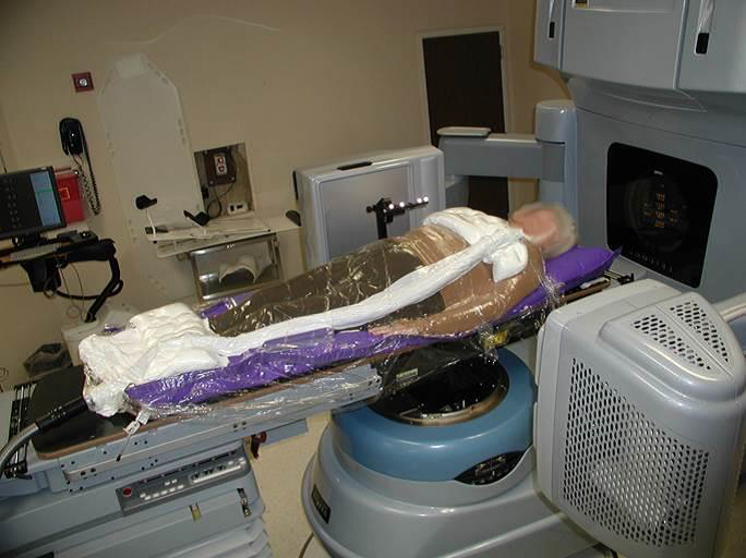
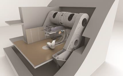
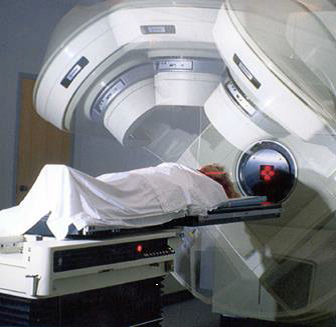 Fig 4:
Stereotactic Body Radiotherapy.
Uses CT, PET or MRI scans to create a 3-D picture of the tumor and surrounding anatomy.Improved precision, decreased normal tissue damage.
For patients treated with 3-D or IMRT. Physicians use frequent imaging of the tumor, bony anatomy or implanted fiducial markers for daily set-up accuracy.Imaging performed using CT scans, high quality X-rays, MRI or ultrasound. Motion of tumors can be tracked to maximize tumor coverage and minimize dose to normal tissues.
Uses CT, PET or MRI scans to create a 3-D picture of the tumor and surrounding anatomy.Improved precision, decreased normal tissue damage.
A highly sophisticated form of 3-D CRT allowing radiation to be shaped more exactly to fit the tumor. Radiation is broken into many beamlets, the intensity of each can be adjusted individually. IMRT allows higher doses of radiation to be delivered to the tumor while sparing healthier surrounding tissue.
For patients treated with 3-D or IMRT. Physicians use frequent imaging of the tumor, bony anatomy or implanted fiducial markers for daily set-up accuracy.Imaging performed using CT scans, high quality X-rays, MRI or ultrasound. Motion of tumors can be tracked to maximize tumor coverage and minimize dose to normal tissues.
It uses accurately target tumors by adjusting for tumor motion during intensity modulated radiation therapy (IMRT). Tumors often move as a result of breathing and other involuntary movement in the body. Respiratory gating enables it to paint optimal doses of radiation onto tumors with greater accuracy. The system senses and accounts for motion as the patient breathes in and out, helping us to target the tumor and protect healthy tissues from receiving unnecessary radiation.
RapidArc is an advanced technology use to deliver intensity modulated radiation therapy (IMRT) with speed and precision. RapidArc shortens treatment times to one-half to one-eighth that of conventional radiation therapy. In a single 360-degree rotation, a linear accelerator revolves around the patient, delivering a sculpted, tightly-focused beam of radiation directly to a tumor in less than two minutes. During treatment, the beam is continually shaped and modulated to conform to the shape of the tumor. Treatment can also be delivered from nearly any angle. This results in better tumor targeting and less damage to surrounding healthy tissue.
SRS is a specialized type of external beam radiation that uses focused radiation beams targeting a well-defined tumor.SRS relies on detailed imaging, 3-D treatment planning and complex immobilization for precise treatment set-up to deliver the dose with extreme accuracy. It is used on the brain or spine. Typically delivered in a single treatment or fraction.
SBRT refers to stereotactic radiation treatments in 1-5 fractions on specialized linear accelerators. It uses sophisticated imaging, treatment planning and immobilization techniques. Respiratory gating may be necessary for motions management, e.g., lung tumors. SBRT is used for a number of sites: spine, lung, liver, brain, adrenals, pancreas and prostate.
Protons are charged particles that deposit most of their energy at a given depth, minimizing risk to tissues beyond that point. Allows for highly specific targeting of tumors located near critical structures. Increasingly available in the U.S.Most commonly used in treatment of pediatric, CNS and intraocular malignancies. Data maturing for use in other tumor sites.
Intracavitary implants: Radioactive sources are placed in a cavity near the tumor (breast, cervix, uterine).Interstitial implants: Sources placed directly into the tissue (prostate, vagina).Intra-operative implants: Surface applicator is in direct contact with the surgical tumor bed.
Low-Dose-Rate (LDR): Radiation delivered over days and months. Prostate, breast, head and neck, and gynecologic cancers may be treated with LDR brachytherapy. High-Dose-Rate (HDR): High energy source delivers the dose in a matter of minutes rather than days. Gynecologic, breast, head and neck, lung, skin and some prostate implants may use HDR brachytherapy.
IORT delivers a concentrated dose of radiation therapy to a tumor bed during surgery. Advantages: Decrease volume of tissue in boost field, Increase the effective dose.Multiple sites: Breast, Pancreas, stomach, lung, esophagus, colorectal, sarcomas, pediatric tumors, bladder, kidney, gyn.
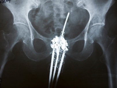 Fig :
Fig : Combo Brachytherapy Improves Local Control in Cervical Cancer